

Wrist Injuries & Conditions
Your wrist is made up of eight small bones (carpal bones) plus two long bones in your forearm — the radius and the ulna. The most commonly injured carpal bone is the scaphoid bone, located near the base of your thumb. The carpal bones are small square, oval, and triangular bones. The cluster of carpal bones in the wrist make it both strong and flexible. Your wrist and hand wouldn’t work the same if the wrist joint was only made up of one or two larger bones.
At Aptiva Health, we offer same-day and walk-in appointments for elbow injuries and conditions to evaluate, diagnose, and make the appropriate referral for additional treatment based upon your specific elbow injury or condition. We treat these conditions in our General Medicine, Orthopedics, Sports Medicine, and Physical Therapy departments.
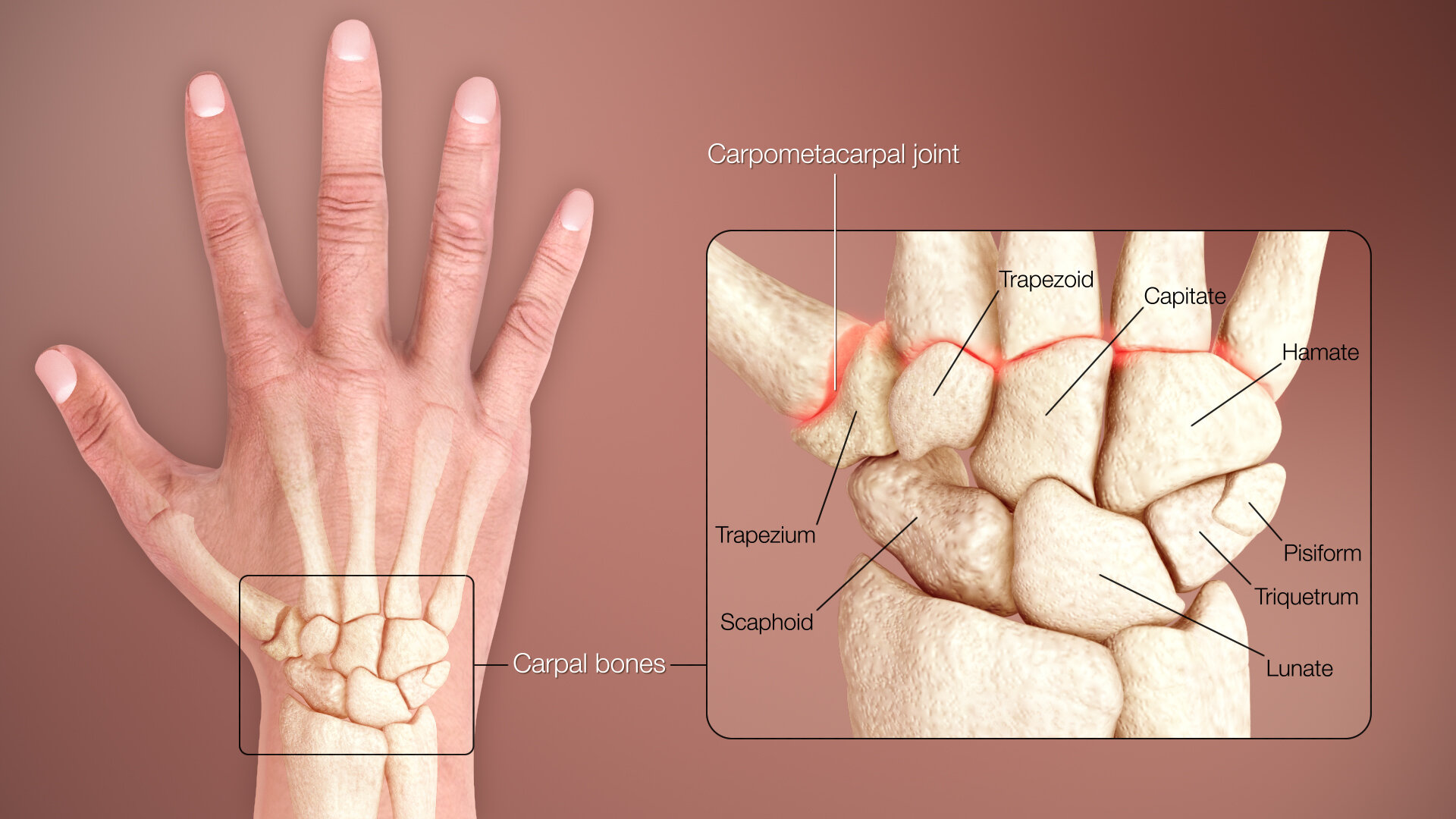
The eight carpal bones are:
Scaphoid: long boat-shaped bone under your thumb
Lunate: a crescent-shaped bone beside the scaphoid
Trapezium: a rounded-square shaped bone above the scaphoid and under the thumb
Trapezoid: bone beside the trapezium that’s shaped like a wedge
Capitate: an oval or head-shaped bone in the middle of the wrist
Hamate: bone under the pinky finger side of the hand
Triquetrum: pyramid-shaped bone under the hamate
Pisiform: a small, round bone that sits on top of the triquetrum

Anatomy of the Hand
The wrist has three main joints. This makes the wrist more stable than if it had only one joint. It also gives your wrist and hand a wide range of movement. The wrist joints let your wrist move your hand up and down, like when you lift your hand to wave. These joints allow you to bend your wrist forward and backward, side to side, and to rotate your hand.
Radiocarpal joint
This is where the radius — the thicker forearm bone — connects with the bottom row of wrist bones: the scaphoid, lunate and triquetrum bones. This joint is mainly on the thumb side of your wrist.
Ulnocarpal joint
This is the joint between the ulna — the thinner forearm bone — and the lunate and triquetrum wrist bones. This is the pinky finger side of your wrist.
Distal radioulnar joint
This joint is in the wrist but doesn’t include the wrist bones. It connects the bottom ends of the radius and ulna.
Common Wrist Injuries & Conditions
There are a number of injuries that may occur in the hands or wrists. They can be classified into two main categories:
Traumatic (acute) and
Overuse (chronic).

Traumatic injuries are more likely to occur in persons who participate in sports that require higher levels of contact (i.e., football, hockey, or wrestling), whereas overuse injuries result in people that participate in jobs or activities that require them to “overdo” a particular movement (i.e., tennis, typing, factory line work).
Some common traumatic injuries include joint dislocations, sprains, muscle strains, broken bones, tendon inflammation, and ligament tears.

Overuse injuries are stress-induced and include tendon inflammation and dislocation, nerve injury, and over use stress fractures. Long-term disability is less likely to occur from overuse injuries than from traumatic injuries. However, if left untreated, overuse injuries of the wrist can interfere with activities of daily living and cause ongoing pain and issues if left untreated. Surgical treatment may be required if an overuse injury persists.
Some of the more common types of wrist injuries and disorders are:

Carpal Tunnel Syndrome, also called median nerve compression, is a condition that causes numbness, tingling, or weakness in your hand. It happens because of pressure on your median nerve, which runs the length of your arm, goes through a passage in your wrist called the carpal tunnel, and ends in your hand.

Ganglion Cysts are lumps that most commonly develop in the wrist. They're typically round or oval and are filled with a jelly-like fluid. Ganglion cysts are noncancerous lumps that most commonly develop along the tendons or joints of your wrists or hands. They also may occur in the ankles and feet.

Wrist Fracture is a medical term for a broken wrist. The wrist is made up of eight small bones which connect with the two long forearm bones called the radius and ulna. Although a broken wrist can happen in any of these 10 bones, by far the most common bone to break is the radius.

Wrist Osteoarthritis, the most common type of arthritis. It is caused by wear and tear of the joints. As with any joint affected by osteoarthritis, the primary symptom associated with wrist osteoarthritis is pain. In the early stages of osteoarthritis, pain is brought on by activity.

Wrist Sprain occurs when the strong ligaments that support the wrist stretch beyond their limits or tear. This occurs when the wrist is bent or twisted forcefully, such as caused by a fall onto an outstretched hand. Wrist sprains are common injuries. They can range from mild to severe depending on how much damage there is to the ligaments.

Wrist Tendonitis, also called tenosynovitis, is a common condition characterized by irritation and inflammation of the tendons around the wrist joint. Many tendons surround the wrist joint. Wrist tendonitis usually affects one of the tendons, but it may also involve two or more. Often, wrist tendonitis occurs at points where the tendons cross each other or pass over a bony prominence. These are possible sites of irritation and can lead to discomfort when moving the wrist joint.
Diagnosis
Your orthopedic specialist at Aptiva Health will likely begin with a comprehensive physical examination and imaging to assess and accurately diagnose your wrist injury or condition.
Your doctor also might recommend one or more imaging tests to get a closer look:
X-rays. These can help your doctor find bone spurs, arthritis, and other bone-related causes of your wrist pain.
Magnetic resonance imaging (MRI) scan. This uses radio waves and a powerful magnet to make detailed images of your wrist.
Computerized tomography (CT) scan. This is a series of X-rays taken from different angles. When they’re put together, they can give your doctor a better look at what’s happening with your wrist.
Electromyography (EMG). This measures the electrical activity in your muscles to see if there are any problems with your nerves.
Treatment
For dislocations, separations and fractures, your orthopedic specialist will determine whether setting the wrist is the best treatment pathway following a thorough examination or whether surgical intervention is necessitated based on the severity of the dislocation, separation or fracture.

For many other issues that cause wrist pain, your orthopedist may suggest rest, heat or ice and a medicine like ibuprofen or aspirin to reduce the pain and swelling. Additionally, your orthopedist may start you out on a physical therapy regimen to see if symptoms approve.

If your wrist doesn’t improve after these first steps, your orthopedist may try injecting a corticosteroid (an anti-inflammatory medicine) into the joint to relieve swelling and pain. Your orthopedist may also discuss other types of injections such as PRP injections or stem cell injections to treat your wrist pain.

Sometimes carpal tunnel syndrome, ligament tears, wrist fractures, and other wrist injuries and conditions do not improve with rest, physical therapy, or conservative treatment options. In these instances, your orthopedic surgeon may recommend surgical intervention, such as wrist arthroscopy after exhausting conservative treatment measures. Wrist arthroscopy utilizes a small fiber optic instrument called an arthroscope that enables the surgeon to see inside the joint without making large incisions into the muscle and tissue. In a wrist arthroscopy, the surgeon makes small incisions (called portals) through the skin in specific locations around a joint. These incisions are less than half an inch long. The arthroscope, which is approximately the size of a pencil, is inserted through these incisions. The arthroscope contains a small lens, a miniature camera, and a lighting system.
The three-dimensional images of the joint are projected through the camera onto a television monitor. The surgeon watches the monitor as he or she moves the instrument within the joint. Probes, forceps, knives, and shavers at the ends of the arthroscope are used to correct problems uncovered by the surgeon.