
Knee Injuries & Conditions
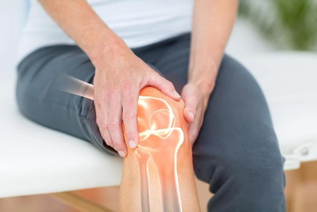
Knee pain is one of the most common reasons people see an orthopedic specialist — and the cause can range from a simple sprain that heals on its own to a complex ligament tear that needs surgical reconstruction. Aptiva Health treats the full continuum of knee injuries and conditions — from acute sports injuries and work-related trauma to chronic arthritis and overuse conditions — across our locations in Louisville, Elizabethtown, Mount Washington, Lexington, Northern Kentucky, and Indianapolis. With on-site X-ray and MRI, same-day and walk-in appointments, an experienced orthopedic and sports medicine team led by Dr. J. Steve Smith (Kerlan-Jobe fellowship), and a complete continuum of conservative through surgical care, we find the source of your knee pain and build a clear treatment plan.
Medically reviewed by J. Steve Smith, MD and the Aptiva Health Orthopedic & Sports Medicine team — June 2026.
Schedule your appointment today!
Knee Problems — The Short Version
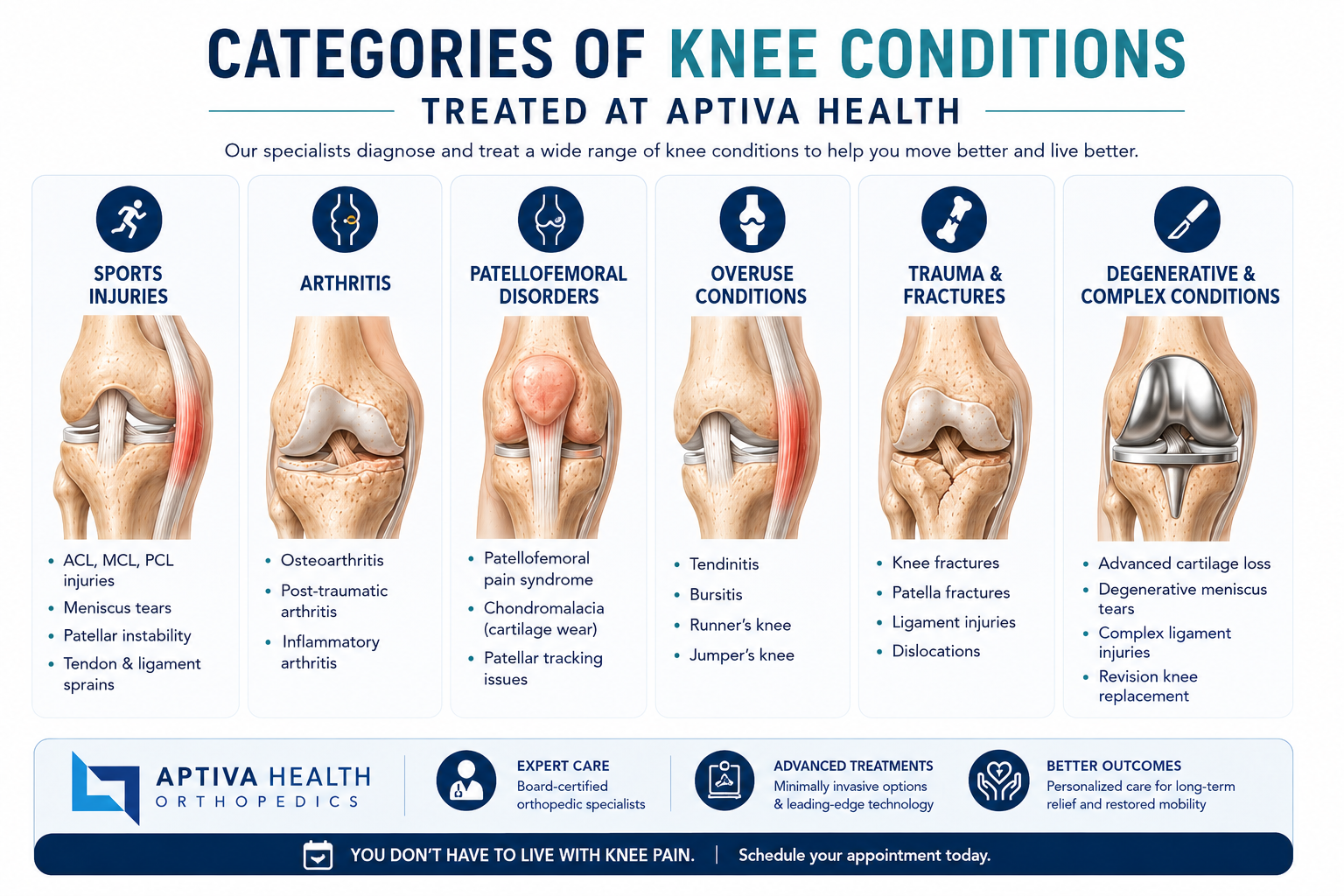
The five categories of knee problems: Ligament injuries, cartilage injuries (meniscus and articular cartilage), joint arthritis, tendon and soft-tissue conditions, and acute traumatic injuries (fractures and dislocations).
Most common knee ligament injury: MCL tear — more common than ACL tear.
Most common knee surgery: Arthroscopic meniscus surgery — approximately 850,000 procedures performed in the United States each year.
Most common cause of chronic knee pain: Knee osteoarthritis.
How most knee injuries are diagnosed: Physical examination plus magnetic resonance imaging (MRI) — Aptiva offers affordable on-site MRI at our locations.
Do most knee injuries require surgery? No. The majority of knee injuries respond to a structured course of conservative care — physical therapy, anti-inflammatory medication, activity modification, bracing, and injections — with surgery reserved for specific situations where conservative care has failed or the injury pattern requires it.
Knee Conditions We Treat
Aptiva Health treats the complete range of knee injuries and conditions across our General Medicine, Orthopedics, Sports Medicine, Pain Management, and Physical Therapy departments. Below are the categories and the specific conditions within each.
Knee Ligament Injuries
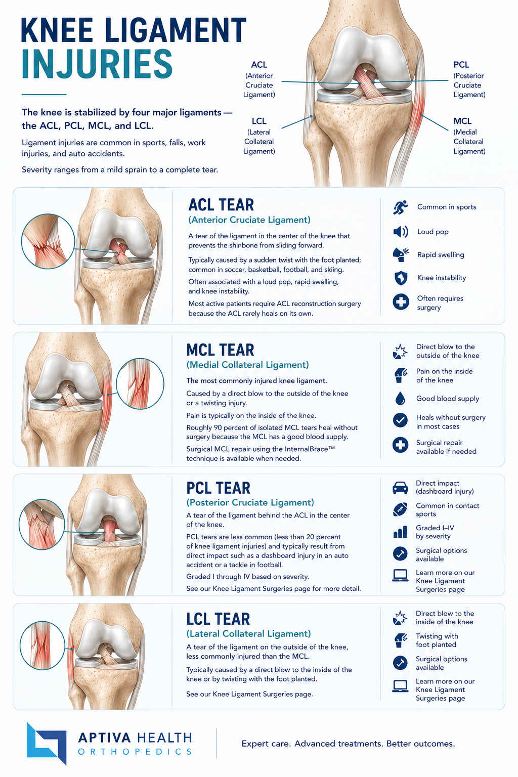
The knee is stabilized by four major ligaments — the ACL, PCL, MCL, and LCL. Ligament injuries are common in sports, falls, work injuries, and auto accidents. Severity ranges from a mild sprain to a complete tear.
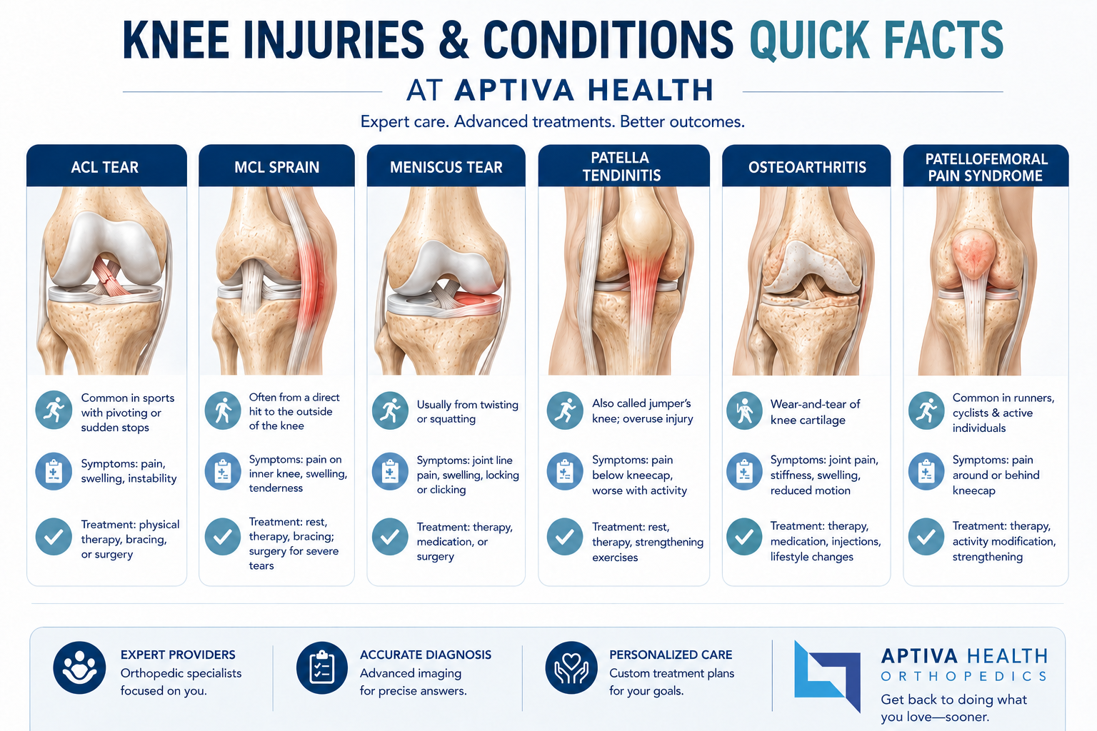
ACL Tear (Anterior Cruciate Ligament) — A tear of the ligament in the center of the knee that prevents the shinbone from sliding forward. Typically caused by a sudden twist with the foot planted; common in soccer, basketball, football, and skiing. Often associated with a loud pop, rapid swelling, and knee instability. Most active patients require ACL reconstruction surgery because the ACL rarely heals on its own.
MCL Tear (Medial Collateral Ligament) — The most commonly injured knee ligament. Caused by a direct blow to the outside of the knee or a twisting injury. Pain is typically on the inside of the knee. Roughly 90 percent of isolated MCL tears heal without surgery because the MCL has a good blood supply. Surgical MCL repair using the InternalBrace™ technique is available when needed.
PCL Tear (Posterior Cruciate Ligament) — A tear of the ligament behind the ACL in the center of the knee. PCL tears are less common (less than 20 percent of knee ligament injuries) and typically result from direct impact such as a dashboard injury in an auto accident or a tackle in football. Graded I through IV based on severity. See our Knee Ligament Surgeries page for more detail.
LCL Tear (Lateral Collateral Ligament) — A tear of the ligament on the outside of the knee, less commonly injured than the MCL. Typically caused by a direct blow to the inside of the knee or by twisting with the foot planted. See our Knee Ligament Surgeries page.
When a knee injury involves more than one ligament — often called a multi-ligament knee injury or, when the ACL, MCL, and medial meniscus are all torn together, the unhappy triad — diagnosis and surgical planning become more complex and benefit from a fellowship-trained sports medicine surgeon.

Knee Cartilage Injuries
The knee contains two types of cartilage: the menisci (C-shaped pads that cushion the joint) and the articular cartilage (the smooth covering on the ends of the bones that allows the joint to glide).
Knee Meniscus Tear — Damage to one of the C-shaped pads that cushion the knee, caused either by sudden twisting (traumatic tear) or by gradual wear (degenerative tear). Most common knee surgery overall. Symptoms include joint line pain, swelling, clicking, catching, and locking. Treatment ranges from physical therapy to arthroscopic meniscus repair or partial meniscectomy.
Articular Cartilage Damage — Damage to the smooth surface cartilage of the femur, tibia, or patella. Causes include acute trauma, repetitive impact, and progression of degenerative joint disease. Treatment options range from physical therapy and bracing to cartilage restoration procedures and, in advanced cases, knee replacement.
Chondromalacia Patellae (Runner's Knee) — Softening and deterioration of the cartilage on the underside of the kneecap, common in young active individuals and in older adults with knee arthritis. Symptoms include anterior knee pain and grinding sensations. Often responds well to physical therapy, activity modification, and quadriceps strengthening.

Knee Arthritis
Arthritis is the most common cause of chronic knee pain and disability. The three most common types affecting the knee are:
Osteoarthritis — The classic age-related "wear and tear" arthritis. Cartilage progressively softens and wears away, causing pain, stiffness, swelling, and reduced motion. Treatment ranges from anti-inflammatory medication, physical therapy, and bracing through corticosteroid and hyaluronic acid injections, PRP therapy, and ultimately partial or total knee replacement for advanced cases.
Rheumatoid Arthritis — An autoimmune inflammatory condition that attacks the joint lining and can damage knee cartilage and bone. Often requires coordinated care with rheumatology in addition to orthopedic management.
Post-Traumatic Arthritis — Arthritis that develops in a knee that was previously injured, often years after a major ligament tear, meniscus tear, or fracture. Can affect younger adults whose knees were injured in sports or accidents in their teens or twenties.
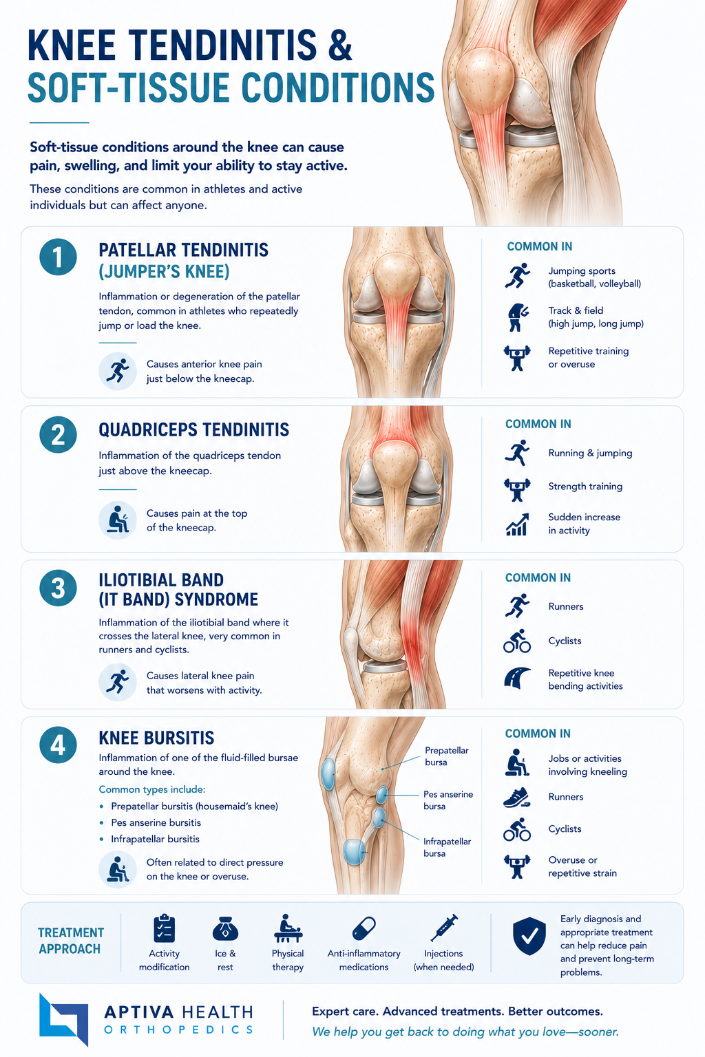
Knee Tendinitis & Soft-Tissue Conditions
Patellar Tendinitis (Jumper's Knee) — Inflammation or degeneration of the patellar tendon, common in athletes who repeatedly jump or load the knee. Causes anterior knee pain just below the kneecap.
Quadriceps Tendinitis — Inflammation of the quadriceps tendon just above the kneecap.
Iliotibial Band (IT Band) Syndrome — Inflammation of the iliotibial band where it crosses the lateral knee, very common in runners and cyclists. Causes lateral knee pain that worsens with activity.
Knee Bursitis — Inflammation of one of the fluid-filled bursae around the knee. Common types include prepatellar bursitis (housemaid's knee), pes anserine bursitis, and infrapatellar bursitis. Often related to direct pressure on the knee or overuse.

Acute Traumatic Knee Injuries
Knee Fractures — Breaks in any of the three bones that form the knee: the femur (especially the femoral condyles), the tibia (especially the tibial plateau), or the patella (kneecap). Tibial plateau fractures are common in auto accidents and falls from height. Aptiva Health offers same-day evaluation, on-site X-ray and MRI, and orthopedic trauma management for non-emergency knee fractures.
Patellar Dislocation — The kneecap slides out of its normal track, typically toward the outside of the knee. Common in adolescents and young adults. First-time dislocations often respond to bracing and physical therapy; recurrent dislocations may require surgical stabilization.
Knee Dislocation — A true knee dislocation, in which the entire joint dislocates (not just the kneecap), is a serious orthopedic emergency that requires immediate evaluation to rule out vascular injury. Inappropriate or delayed treatment may result in loss of the leg.
Mechanical Knee Problems — Including loose bodies in the joint (fragments of cartilage or bone that float freely and cause locking or catching) and meniscal cyst formation.

Knee Procedures We Perform
When conservative care has failed or the injury pattern requires surgical management, Aptiva Health offers the full range of arthroscopic and reconstructive knee procedures.
Knee Meniscus Surgery — Arthroscopic meniscus repair (preserves the meniscus by suturing the tear), arthroscopic partial meniscectomy (trims the torn portion), and meniscal transplant for select younger patients.
ACL Reconstruction — Arthroscopic reconstruction of the anterior cruciate ligament using a graft (typically patellar tendon, hamstring, or quadriceps tendon).
MCL Surgery — MCL repair using the InternalBrace™ technique (a 2 mm FiberTape® suture between two SwiveLock® anchors that internally splints the healing ligament), or MCL reconstruction for chronic instability.
Knee Ligament Surgeries — Comprehensive overview of ACL, PCL, MCL, and LCL surgical approaches, including multi-ligament reconstruction.
Knee Replacement — Partial or total knee arthroplasty for advanced arthritis, with more than 90 percent of modern total knee replacements still functioning well at 15 years.
Knee Arthroscopy — Minimally invasive evaluation and treatment of multiple knee conditions including loose body removal, synovectomy, and cartilage debridement.

When to See a Knee Specialist
Mild knee pain — particularly after exercise or a long day on your feet — often resolves with rest, ice, compression, elevation, and a few days of anti-inflammatory medication. You should see a knee specialist if you experience any of the following:
Knee pain lasting more than two weeks despite rest and over-the-counter pain medication
A pop or tearing sensation at the time of injury
Persistent knee swelling, especially if it returns after activity
Knee instability — a sensation that the knee is buckling or giving way
Knee locking or catching — particularly suggestive of a meniscus tear
Inability to fully straighten or fully bend the knee
Difficulty bearing weight on the affected leg
Knee pain following a sports injury, work injury, or auto accident
Recurrent knee pain or recurrent injury to the same knee
A previous knee surgery and new symptoms in the same or opposite knee
You should seek immediate orthopedic or emergency evaluation for:
A knee dislocation (the entire joint, not just the kneecap)
Sudden severe swelling with inability to bear weight
Numbness, tingling, or cold sensation in the foot below an injured knee
An obvious deformity of the knee or leg
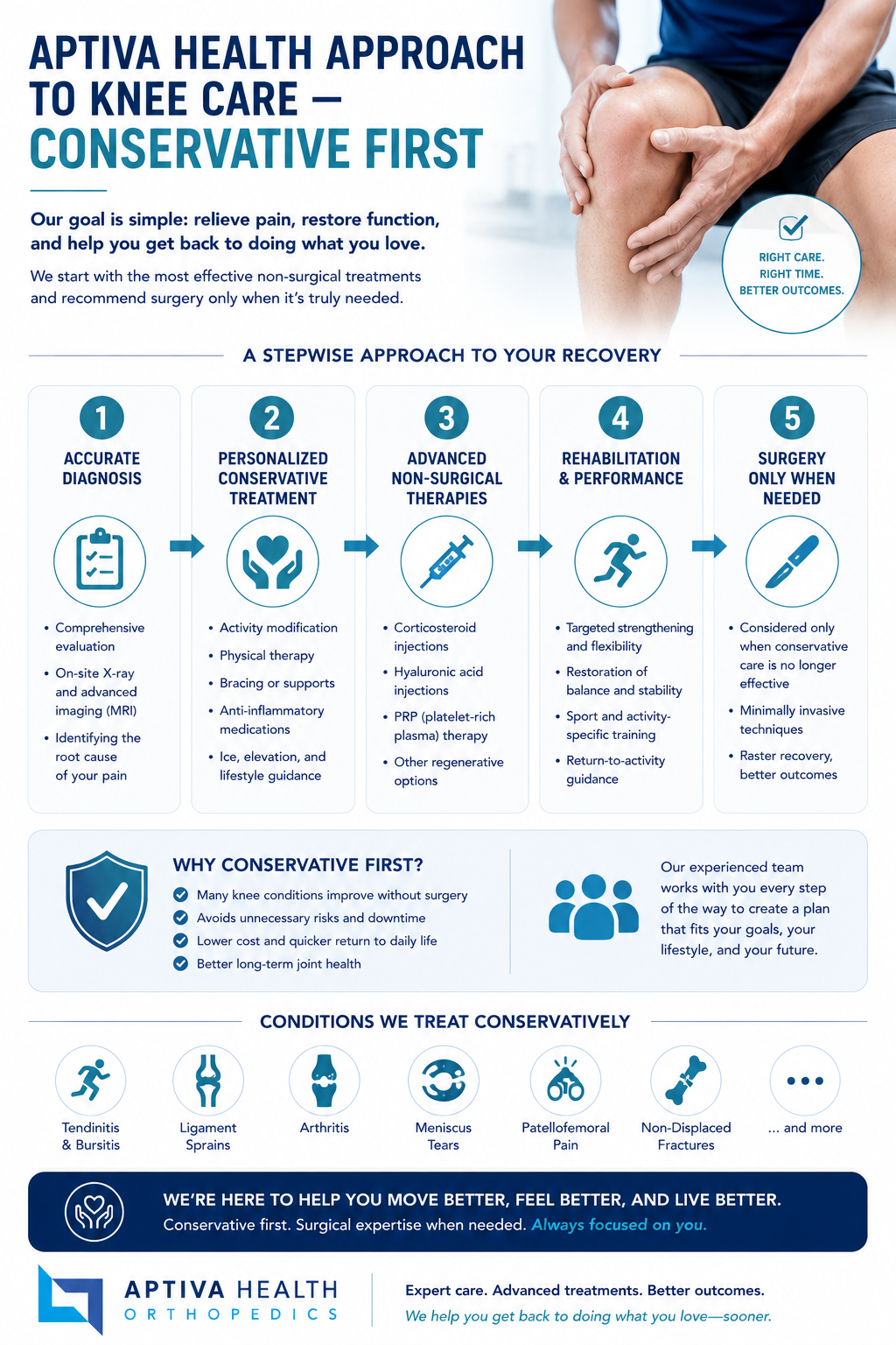
Aptiva's Approach to Knee Care
Diagnosis first. Knee pain has dozens of possible causes, and the right treatment depends entirely on the correct diagnosis. Every Aptiva knee evaluation includes a focused history, a thorough physical exam (including ligament stability tests, meniscus tests, and range-of-motion assessment), and imaging when indicated. On-site X-ray and MRI mean most patients can be evaluated and have imaging completed in a single visit.
Conservative care first. The majority of knee conditions respond to a structured course of non-surgical care. Our toolkit includes:
Orthopedic physical therapy and sports physical therapy — strengthening, range of motion, neuromuscular retraining, and sport- or task-specific rehabilitation
Anti-inflammatory medication
Bracing — hinged braces for ligament protection, sleeves for support, and unloader braces for unicompartmental arthritis
Activity modification and ergonomic guidance
Corticosteroid injections for inflammatory and arthritic pain
Hyaluronic acid (viscosupplementation) injections for knee arthritis
Platelet-rich plasma (PRP) for selected tendon and ligament injuries
Surgery when it's the right answer. When conservative care has been exhausted, when the injury pattern requires surgical management, or when an active patient needs to return to high-demand activity, we offer the full range of arthroscopic and reconstructive knee procedures using modern minimally invasive techniques.
Second opinions welcome. We routinely see patients who have been told they need knee surgery elsewhere and want an independent evaluation before proceeding. Our team will review your imaging, conduct an independent assessment, and discuss whether the recommended procedure is the best option — or whether an alternative might be appropriate.
Your Aptiva Health Knee Care Team
At Aptiva Health, your knee care is delivered by a multidisciplinary team of board-certified orthopedic surgeons, sports medicine specialists, advanced practice providers, and orthopedic physical therapists who care for knee patients every day — from weekend warriors to professional athletes.

Dr. J. Steve Smith — Director of Orthopedic Surgery & Sports Medicine; completed his sports medicine fellowship at the Kerlan-Jobe Orthopaedic Clinic in Los Angeles — the same program that trains team physicians for the LA Dodgers, Lakers, Kings, and Angels. Has served on the medical staffs of the Los Angeles Lakers, Los Angeles Dodgers, and Anaheim Ducks.
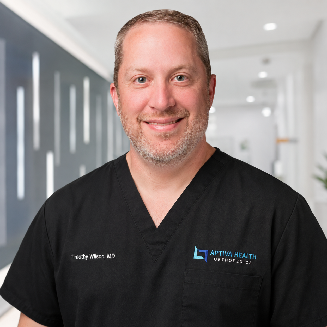
Dr. Timothy Wilson — Board-certified orthopedic surgeon with subspecialty certification in Sports Medicine from the American Board of Orthopaedic Surgery. More than 20 years caring for athletes across Kentucky, with past appointments as team physician for the University of Kentucky Athletics, Kentucky State University, Morehead State University, Georgetown College, and Scott County High School.

Dr. Shawn Price — Board-certified orthopedic surgeon; treats the full range of knee conditions including arthritis, joint replacement, bone sarcoma, meniscus tears, and knee ligament tears.

Dr. D. Philip Stickney — Board-certified orthopedic surgeon since 2002; treats the full range of knee conditions including meniscus tears, ligament injuries, and arthritis at Aptiva's Northern Kentucky (Hebron) and Indianapolis locations.
Advanced Practice Providers
Michael Gilbert, PA-C — Orthopedic physician assistant for 30 years. Provides same-week new-patient evaluations, conservative-care coordination, knee injections, bracing fittings, and post-operative follow-up.
Bradley Stephenson, PA-C — Orthopedic physician assistant. Provides new-patient evaluations, conservative-care coordination, knee injections, and post-operative follow-up for the Aptiva Health orthopedic and sports medicine team. (Verify Squarespace slug before adding hyperlink — bio page URL pending.)
Bryan Davidson, PA-C — Orthopedic physician assistant. Treats orthopedic and sports medicine patients across the Aptiva Health network. (Verify Squarespace slug before adding hyperlink — bio page URL pending.)
Becky Kostyo, APRN — Orthopedic nurse practitioner. Works directly with the Aptiva Health knee team to evaluate, diagnose, and manage knee conditions including conservative care, knee injections, and pre- and post-operative coordination.
Physical Therapy & Imaging
Aptiva's orthopedic physical therapy, sports physical therapy, and imaging services are integrated with the knee team so your evaluation, MRI, bracing, injection, and treatment all happen under one roof.
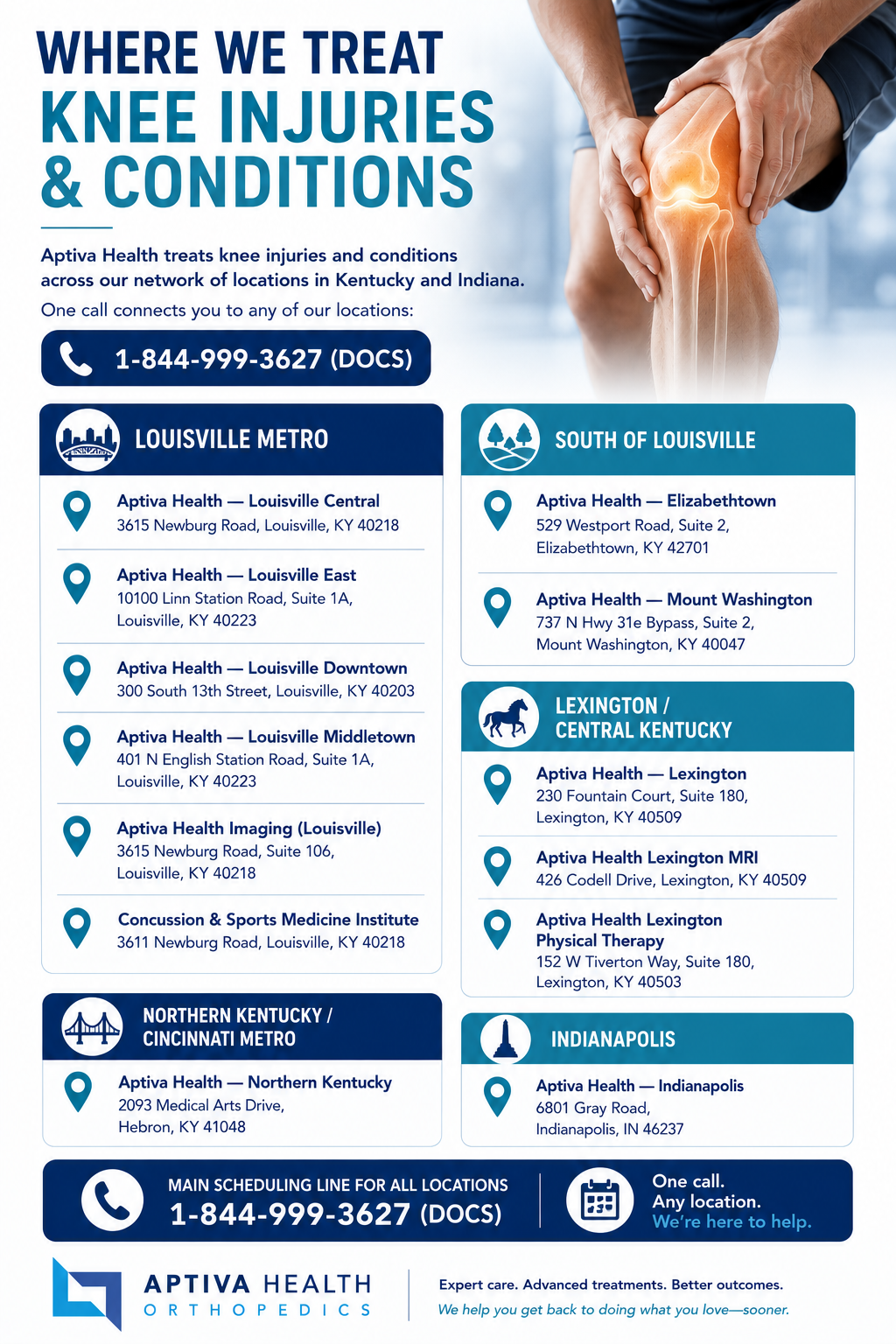
Where We Treat Knee Injuries & Conditions
Aptiva Health treats knee injuries and conditions across our network of locations in Kentucky and Indiana. One call connects you to any of our locations: 1-844-999-3627 (DOCS).
Louisville Metro
Aptiva Health — Louisville Central — 3615 Newburg Road, Louisville, KY 40218
Aptiva Health — Louisville East — 10100 Linn Station Road, Suite 1A, Louisville, KY 40223
Aptiva Health — Louisville Downtown — 300 South 13th Street, Louisville, KY 40203
Aptiva Health — Louisville Middletown — 401 N English Station Road, Suite 1A, Louisville, KY 40223
Aptiva Health Imaging (Louisville) — 3615 Newburg Road, Suite 106, Louisville, KY 40218
Concussion & Sports Medicine Institute — 3611 Newburg Road, Louisville, KY 40218
South of Louisville
Lexington / Central Kentucky
Aptiva Health — Lexington — 230 Fountain Court, Suite 180, Lexington, KY 40509
Aptiva Health Lexington MRI — 426 Codell Drive, Lexington, KY 40509
Aptiva Health Lexington Physical Therapy — 152 W Tiverton Way, Suite 180, Lexington, KY 40503
Northern Kentucky / Cincinnati Metro
Indianapolis
Main scheduling line for all locations: 1-844-999-3627 (DOCS)

Why Choose Aptiva Health for Your Knee Care
Sports medicine pedigree. Dr. J. Steve Smith is fellowship-trained at the Kerlan-Jobe Institute — the program that trains team physicians for the Los Angeles Dodgers, Lakers, Kings, and Angels. Dr. Timothy Wilson carries the American Board of Orthopaedic Surgery's subspecialty certification in Sports Medicine and has served as team physician for the University of Kentucky and multiple Kentucky colleges and high schools.
Diagnosis under one roof. From your first call to a clear diagnosis with imaging, bracing, and a treatment plan — most Aptiva patients can complete their evaluation in a single visit. On-site X-ray and MRI are available at our locations.
Conservative care first. Most knee injuries can be managed without surgery. We don't push surgery — we recommend the least invasive option that will get you back to your sport, your job, and your life.
Modern surgical techniques when surgery is the right answer. When surgery is needed, Aptiva uses arthroscopic and minimally invasive approaches, including the MCL InternalBrace™ technique, anatomical ACL reconstruction, meniscus repair (when feasible) rather than meniscus removal, and modern partial and total knee replacement.
Same-day and walk-in appointments. Most patients see a knee specialist within days — and walk-in immediate injury care is available across our locations.
A complete continuum under one roof. From on-site X-ray and MRI to physical therapy, bracing, injections, PRP therapy, and surgery — we offer the complete knee care continuum so you're not bounced between unrelated providers and facilities.
Multiple convenient locations. Care is available across Louisville, Elizabethtown, Mount Washington, Lexington, Northern Kentucky, and Indianapolis.
Direct access — no referral needed. Schedule directly without a physician referral. We welcome second-opinion patients, including those who have been told elsewhere they need knee surgery.
Transparent insurance and cash-pay pricing. We accept most major insurance, Medicare, Medicaid Managed Care, workers' compensation, and auto insurance (PIP and Medpay). For self-pay patients, transparent bundled pricing and affordable cash-pay MRI are available.
Documentation that supports your case. For knee injuries from work or auto accidents, our team works directly with employers, adjusters, and sports teams on documentation and coordination of care.
Schedule your appointment today!
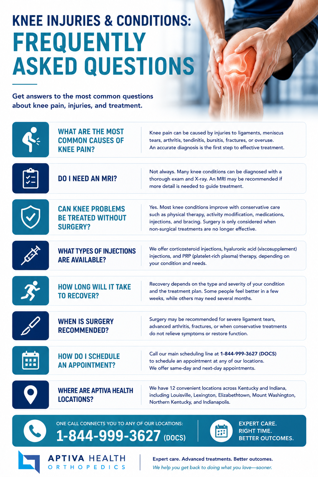
Knee Injuries & Conditions: Frequently Asked Questions
What are the most common knee injuries?
The most common knee injuries fall into four broad categories. Ligament injuries include ACL tears, MCL tears, PCL tears, and LCL tears — the MCL is actually the most commonly injured knee ligament. Cartilage injuries include meniscus tears (approximately 850,000 meniscus procedures are performed in the United States each year) and articular cartilage damage. Joint conditions include knee osteoarthritis, rheumatoid arthritis, and post-traumatic arthritis. Soft tissue and tendon problems include patellar tendinitis (jumper's knee), chondromalacia (runner's knee), iliotibial band syndrome, and bursitis. Acute traumatic injuries include knee fractures and patellar dislocations.
When should I see a knee specialist?
You should see a knee specialist if you have knee pain that does not improve within one to two weeks of rest, ice, and over-the-counter medication, knee swelling that does not resolve, knee instability or a sensation that the knee is giving way, a popping or tearing sensation at the time of injury, knee locking or catching, inability to fully straighten or bend the knee, difficulty bearing weight, or any knee injury sustained during sports, work, or an auto accident. At Aptiva Health, same-day and walk-in knee evaluations are available across all locations.
Do I need a referral to see a knee doctor at Aptiva Health?
No. You can schedule directly with Aptiva Health without a physician referral. We also welcome patients seeking a second opinion, including those who have been told they need knee surgery elsewhere and want an independent evaluation before proceeding.
Should I go to urgent care or an orthopedic specialist for knee pain?
Urgent care is appropriate when you need a fracture ruled out and the only available service is a basic X-ray with general medical evaluation. An orthopedic or sports medicine specialist is the better choice when you have an injury that likely involves a ligament, meniscus, or cartilage; when symptoms have persisted or recurred; when you need MRI; or when you may need physical therapy, injection, or surgical treatment. Aptiva Health combines both — same-day immediate injury evaluation by an orthopedic or sports medicine provider, with on-site X-ray, MRI, and physical therapy — so most patients can be evaluated and start treatment in a single visit.
How does Aptiva Health diagnose knee injuries?
Knee evaluation at Aptiva Health begins with a focused history of how the injury occurred, where the pain is located, and what makes it better or worse. A thorough physical exam evaluates range of motion, joint line tenderness, ligament stability (valgus and varus stress tests, Lachman, anterior and posterior drawer), meniscus tests (McMurray, Apley, Thessaly), and signs of effusion. X-rays are obtained when needed to rule out fracture or assess for arthritis. Magnetic resonance imaging (MRI) is the gold standard for evaluating soft tissue injuries to ligaments, menisci, and cartilage — and Aptiva offers affordable on-site MRI across locations.
Does Aptiva Health offer same-day knee appointments?
Yes. Aptiva Health offers same-day and walk-in appointments for knee injuries and conditions across all of our locations in Louisville, Elizabethtown, Mount Washington, Lexington, Northern Kentucky (Hebron), and Indianapolis. Same-day appointments are most commonly available for acute injuries from sports, work, or auto accidents. Call 1-844-999-3627 (DOCS) to schedule.
Does Aptiva treat sports-related, work-related, and auto accident knee injuries?
Yes. Aptiva Health treats knee injuries from sports, work, and auto accidents, and our team works directly with employers, adjusters, and sports teams on documentation and coordination of care. We accept workers' compensation, auto insurance (PIP and Medpay), and major commercial insurance. Same-day immediate injury evaluation is available.
What insurance does Aptiva Health accept for knee care?
Aptiva Health accepts most major insurance plans, Medicare, Medicaid Managed Care, workers' compensation, and auto insurance (PIP and Medpay). Coverage typically includes office visits, X-ray, MRI, physical therapy, knee injections, and knee surgery when needed. Our team verifies benefits before any visit or procedure and can also provide cash-pay pricing for patients without insurance or with high-deductible plans.
What happens at my first knee appointment at Aptiva?
Your first knee appointment typically includes a focused history about how the injury occurred and what makes the symptoms worse, a thorough physical examination of the knee and surrounding joints, on-site X-rays if needed to rule out fracture or assess for arthritis, a discussion of likely diagnoses and next steps, and a treatment plan that may include physical therapy, bracing, anti-inflammatory medication, injections, MRI, or surgery referral. Many patients leave the first appointment with a clear diagnosis and the start of a treatment plan in hand.
Can I get a second opinion on knee surgery at Aptiva?
Yes. Aptiva Health welcomes patients seeking a second opinion, including those who have been told they need knee surgery elsewhere. Our team will review your imaging, conduct an independent evaluation, and discuss whether the recommended procedure is the best option — or whether an alternative such as continued physical therapy, an interventional pain procedure, or a less invasive surgical approach might be appropriate.
Sources: Mayo Clinic and AAOS OrthoInfo.