

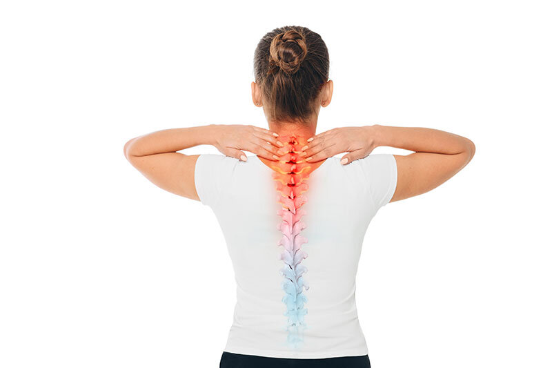
Degenerative Disc Disease
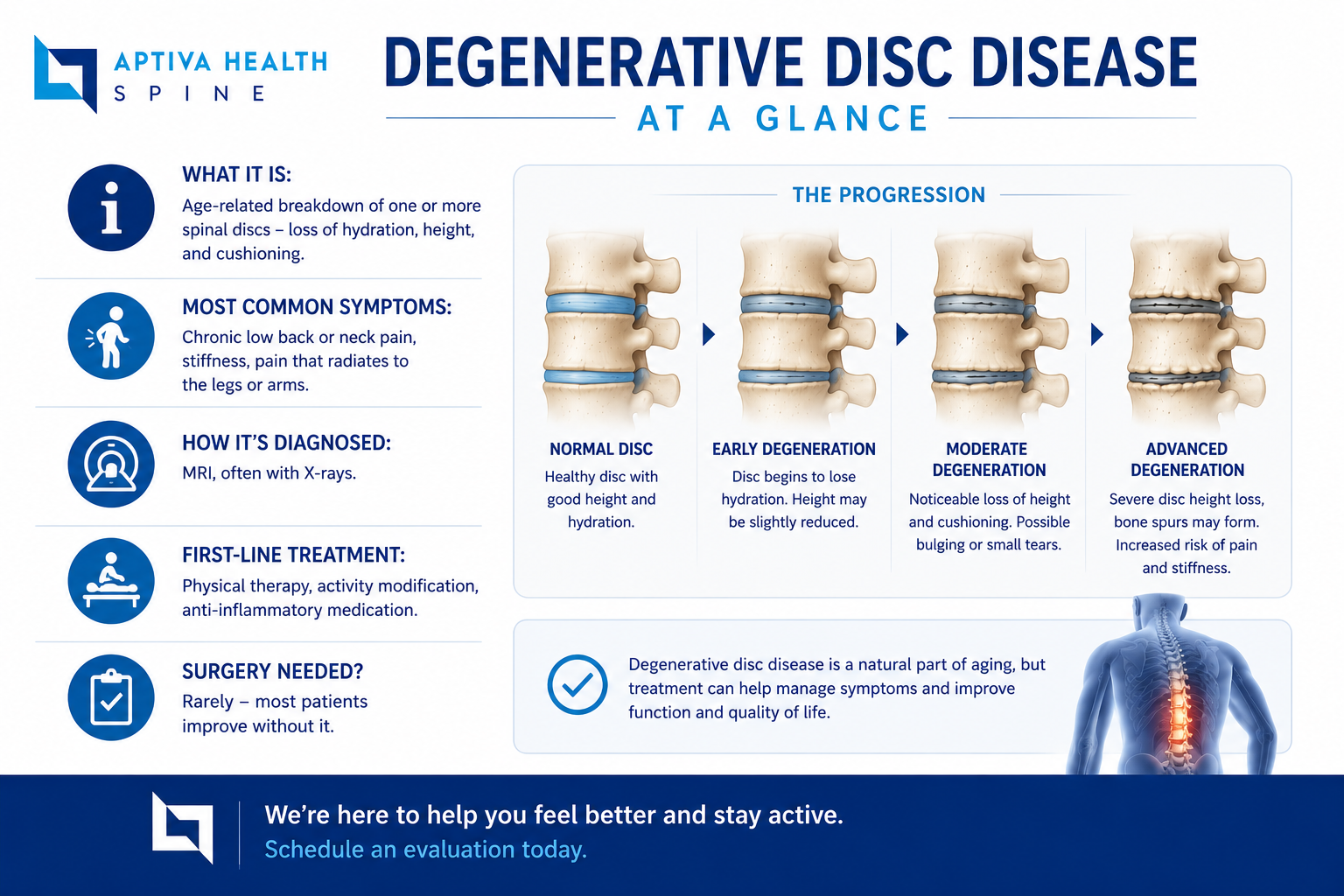
Degenerative disc disease (DDD) is a condition where one or more discs in the back lose their strength and break down. Degenerative disc disease, despite the name, isn’t technically a disease. DDD is a progressive condition that can occur over time from wear and tear, or from injury.
The discs in your back are located in between the vertebrae of the spine. They act as cushions and shock absorbers. Discs help you stand up straight. And they also help you move through everyday motions, such as twisting around and bending over. Some people can have DDD without many symptoms, while others can suffer mild to extreme pain that can interfere with activities of daily living.
Medically reviewed by Michael Casnellie, MD, David McConda, MD, and Jaideep Chunduri, MD on July 24, 2026.
Schedule your appointment today!
What Happens to a Disc as It Degenerates
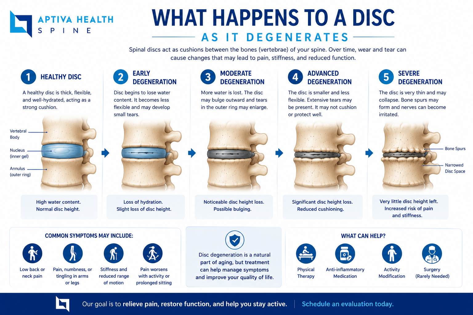
The discs between your vertebrae act as cushions and shock absorbers. Each disc has a tough, fibrous outer ring (the annulus fibrosus) and a soft, gel-like center (the nucleus pulposus). In a healthy young adult, discs are up to 90 percent water.
Two changes drive disc degeneration:
Loss of fluid (disc desiccation). With age, the disc's water content decreases, making the disc thinner and less effective as a shock absorber. This dehydration is one of the earliest visible signs of DDD on MRI - so early, in fact, that radiologists give it its own name: disc desiccation. The space between vertebrae narrows as discs lose height.
Damage to disc structure. Small tears or cracks develop in the outer ring of the disc. The gel-like inner material can push through these tears, resulting in a bulging or herniated disc.
As discs thin, the spine becomes less stable. The body compensates by building bone spurs (osteophytes) along the edges of the vertebrae. These bony projections can press against the spinal cord or nerve roots, causing pain, weakness, and numbness. Over time, disc degeneration can also contribute to spinal stenosis (narrowing of the spinal canal), spondylosis, and radiculopathy.
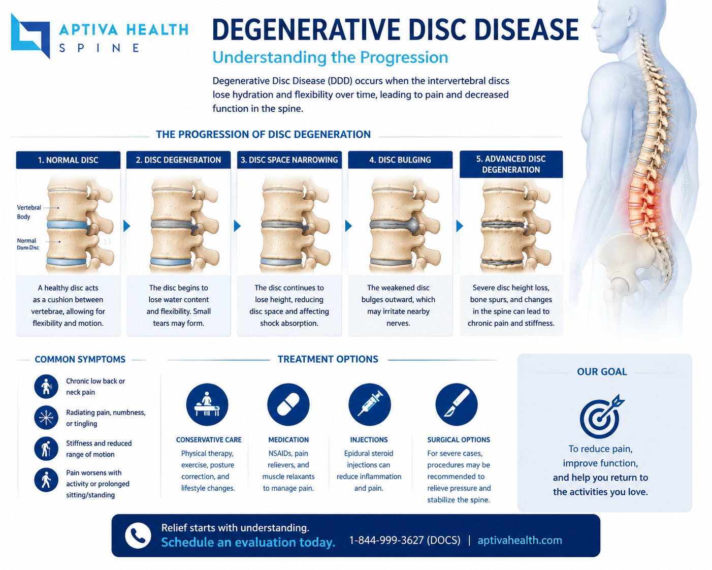
This graphic depicts the difference between a normal disc and a degenerated disc.
Symptoms of Degenerative Disc Disease
Disc degeneration may cause no symptoms at all, or it may cause pain intense enough to interfere with daily activities. Symptoms depend largely on where the degeneration occurs:
Lumbar spine (low back): Chronic low back pain that may radiate to the buttocks and upper thighs; tingling or numbness in the legs or feet. Pain is often worse with sitting, bending, or lifting, and improves with walking or changing position.
Cervical spine (neck): Neck pain that may spread to the shoulder, arm, or hand, sometimes with tingling or numbness.
Instability symptoms: Muscle spasms in the lower back or neck as the body attempts to stabilize the affected segment.
Seek prompt evaluation if you develop progressive leg weakness, numbness in the groin area, or loss of bowel or bladder control - these can signal serious nerve compression that requires urgent treatment.
causes
Intervertebral discs, also known as intervertebral fibrocartilage or spinal discs, provide the padding between the vertebrae of the spine. They have an elastic structure, made of fibrocartilage tissue. The outer part of the disc is known as the annulus fibrosus. It is tough and fibrous, and it consists of several overlapping layers. The inner core of the disc is the nucleus pulposus. It is soft and gelatinous. The intervertebral discs cushion when the spine moves or bears weight. They also help the spine to bend. As people age, repeated daily stresses on the spine and occasional injuries, including minor, unnoticed ones, can damage the discs in the back.
Changes include:
Loss of fluid: The intervertebral discs of a healthy young adult consist of up to 90 percent fluid. With age, the fluid content decreases, making the disc thinner. The distance between vertebrae becomes smaller, and it becomes less effective as a cushion, or shock-absorber.
Disc structure: Very small tears or cracks develop in the outer layer of the disc. The soft and gelatinous material in the inner part may seep through the cracks or tears, resulting in a bulging or rupturing disc. The disc may break into fragments.
When the vertebrae have less padding between them, the spine becomes less stable. To compensate, the body builds osteophytes (bone spurs) small bony projections that develop along the edge of bones. These projections can press against the spinal cord or spinal nerve roots. The development of osteophytes can undermine nerve function and cause pain.
Other problems include:
a breakdown of cartilage, the tissue that cushions the joints
a bulging disc, known as a herniated disc
a narrowing of the spinal canal, or spinal stenosis
These changes can affect the nerves, leading to pain, weakness, and numbness.
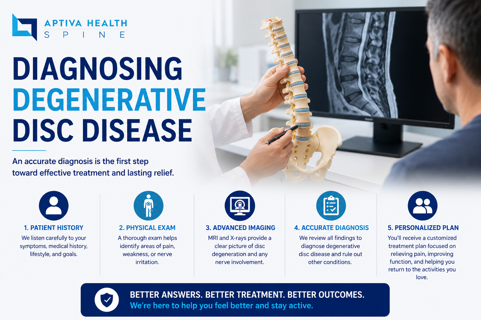
How Degenerative Disc Disease Is Diagnosed
MRI is the most common and most accurate imaging test for DDD. It shows disc hydration, disc height, nerve compression, and the condition of the spinal cord and nerve roots. X-rays are often added to evaluate vertebral alignment, disc space narrowing, and bone spurs - though DDD is not always visible on plain X-rays.
CT scans, myelograms, and EMG/nerve conduction studies are used only in select circumstances.
Aptiva Health operates on-site imaging centers, including cash-pay MRI with transparent pricing (approximately $350) for patients paying out of pocket - no referral required.
X-ray: Application of radiation to produce a film or picture of a part of the body can show the structure of the vertebrae and the outline of the joints. X-rays of the spine are obtained to search for other potential causes of pain, i.e. tumors, infections, fractures, etc.
Computed tomography scan (CT or CAT scan): A diagnostic image created after a computer reads X-rays; can show the shape and size of the spinal canal, its contents and the structures around it.
Magnetic resonance imaging (MRI): A diagnostic test that produces 3D images of body structures using powerful magnets and computer technology; can show the spinal cord, nerve roots and surrounding areas as well as enlargement, degeneration and tumors.
Myelogram: An X-ray of the spinal canal following injection of a contrast material into the surrounding cerebrospinal fluid spaces; can show pressure on the spinal cord or nerves due to herniated discs, bone spurs or tumors.
Electromyogram and Nerve Conduction Studies (EMG/NCS): These tests measure the electrical impulse along nerve roots, peripheral nerves and muscle tissue. This will indicate whether there is ongoing nerve damage, if the nerves are in a state of healing from a past injury or whether there is another site of nerve compression. This test is infrequently ordered.
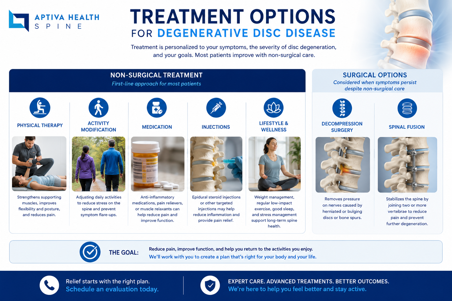
Treatment for Degenerative Disc Disease
Conservative (Non-Surgical) Treatment
Initial treatment for DDD is conservative in nearly all cases:
Physical therapy: A structured program of stretching, strengthening, and core stabilization designed for your specific level and pattern of degeneration. Therapy may also include traction, manual therapy, ice and heat, and electrical muscle stimulation.
Medication: Nonsteroidal anti-inflammatory drugs (NSAIDs) for mild to moderate pain; muscle relaxants when spasms are present.
Activity modification: Adjusting lifting mechanics, posture, and repetitive activities that aggravate symptoms.
Interventional Pain Management
If pain persists despite therapy, an epidural steroid injection delivers anti-inflammatory medication directly to the affected disc level under X-ray (fluoroscopic) guidance. Our interventional pain team, led by Dr. Steven Ganzel, DO, offers the full range of image-guided spine injections.
Surgery
Surgery is considered only when conservative treatment fails and symptoms significantly limit daily life, or when progressive neurological deficits develop. A patient may be a surgical candidate if:
Radicular pain limits normal activity or impairs quality of life
Progressive neurological deficits develop, such as leg weakness or numbness
Loss of normal bowel or bladder function occurs
Standing or walking becomes difficult
Medication and physical therapy are ineffective
Common procedures for DDD of the lumbar spine (low back):
Lumbar laminectomy - removal of all or part of the lamina to relieve pressure on the spinal cord and nerve roots when DDD causes central spinal stenosis
Lumbar discectomy - minimally invasive removal of the damaged portion of a disc to relieve nerve pressure
MAS TLIF (Minimally Invasive Lumbar Interbody Fusion) - a muscle-splitting, minimal-access fusion that stabilizes the affected segment with less blood loss, smaller incisions, and faster recovery than traditional fusion
Common procedures for DDD of the cervical spine (neck):
Cervical disc arthroplasty (artificial disc replacement) - replaces the damaged disc with a prosthetic disc that preserves motion at the treated level. Dr. Casnellie performed the first cervical disc arthroplasty in Kentucky.
Anterior cervical discectomy and fusion (ACDF) - removes the degenerative disc through a small incision at the front of the neck and fuses the adjacent vertebrae. Patients typically go home the same day. Post-operative instructions.
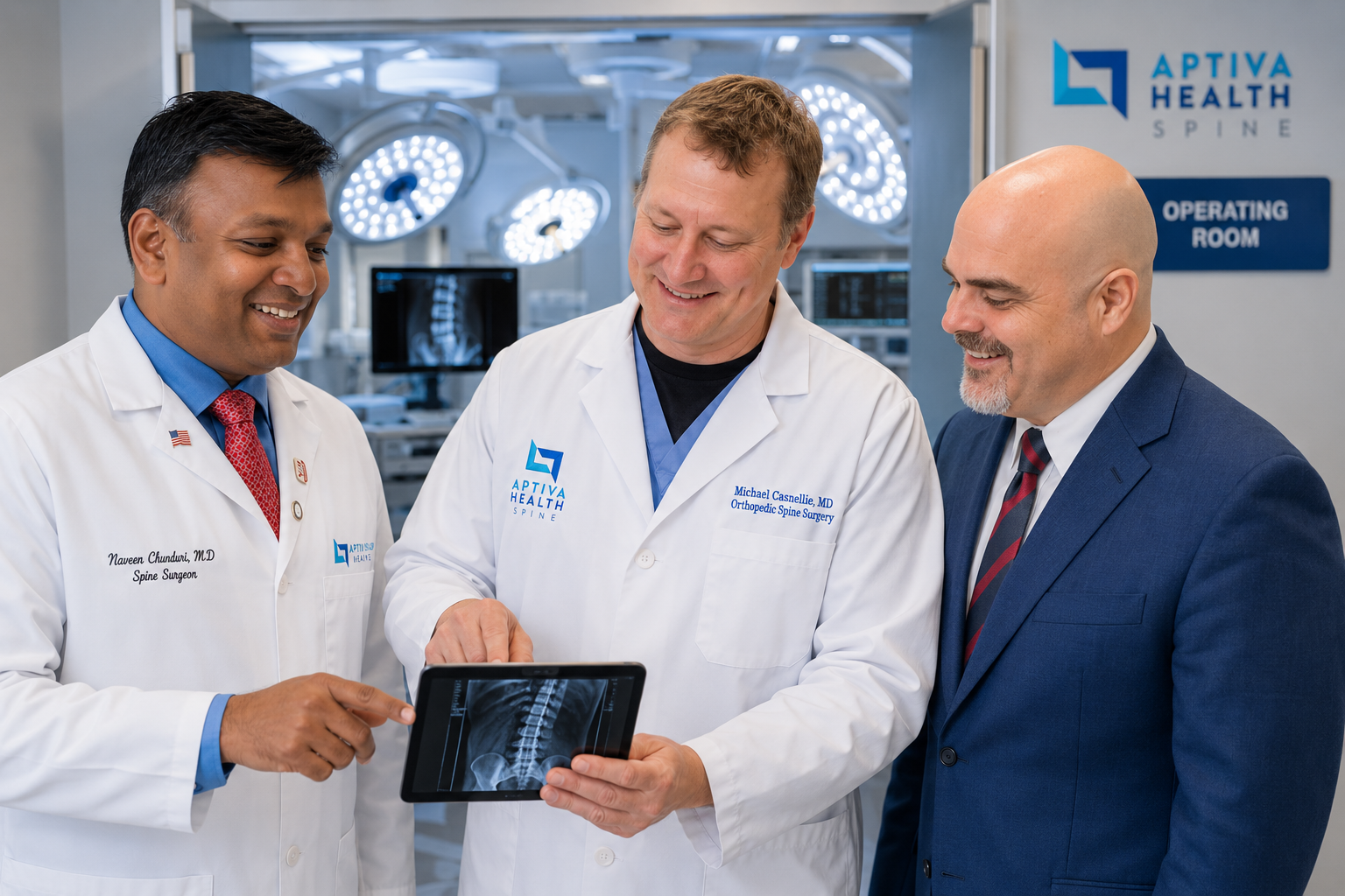
Your Aptiva Health Spine Team
Your care is delivered by a multidisciplinary spine team:
Michael Casnellie, MD - Orthopedic Spine Surgeon
David McConda, MD - Orthopedic Spine Surgeon
Jaideep Chunduri, MD - Orthopedic Spine Surgeon
Steven Ganzel, DO - Interventional Pain Management, double board-certified in PM&R and Interventional Pain
Kayla Troutman, PA-C - Spine Team
David Koonce, DNP - Spine Team
Michael Gilbert, PA-C - Spine Team
Bradley Stephenson, PA-C - Spine Team

Why Patients Choose Aptiva Health
No referral required - schedule directly or walk in
Same-day and walk-in appointments through our Immediate Injury Care clinics
Everything under one roof - evaluation, imaging, physical therapy, injections, and surgery within one coordinated team
Cash-pay MRI with transparent pricing (approximately $350)
Locations across Kentucky and Indiana - Louisville (East, Central, Downtown, Middletown), Lexington, Northern Kentucky (Hebron), Elizabethtown, Mt. Washington, and Indianapolis
Frequently Asked Questions
Is degenerative disc disease actually a disease? No. Despite the name, degenerative disc disease is not a disease - it is an age-related condition in which the discs between the vertebrae gradually lose hydration and height. It is extremely common, and many people with disc degeneration on imaging have no symptoms at all.
What is the difference between degenerative disc disease and disc desiccation? Disc desiccation is the loss of water content within a disc and is one of the earliest visible signs of degenerative disc disease on MRI. Degenerative disc disease is the broader process that can follow, including loss of disc height, tears in the outer disc wall, bone spurs, and nerve compression.
Can degenerative disc disease be reversed? Disc degeneration cannot be reversed, but symptoms can be managed very effectively. Most patients improve with conservative care such as physical therapy, activity modification, and anti-inflammatory medication. Injections and minimally invasive surgery are options when symptoms persist.
How is degenerative disc disease diagnosed? MRI is the most common and most accurate imaging test for degenerative disc disease, often supported by X-rays of the spine. Aptiva Health offers on-site MRI at our imaging centers, including cash-pay MRI with transparent pricing for patients without imaging coverage.
When does degenerative disc disease require surgery? Surgery is considered only when conservative treatment fails and pain limits daily activity, or when progressive neurological symptoms develop such as leg weakness, numbness, or loss of bowel or bladder control. Most patients with degenerative disc disease never need surgery.
Do I need a referral to be seen for degenerative disc disease at Aptiva Health? No referral is required. Aptiva Health offers same-day and walk-in spine evaluations at our locations in Louisville, Lexington, Northern Kentucky, Elizabethtown, Mt. Washington, and Indianapolis. Call 1-844-999-3627 to schedule.